Fig. 4.
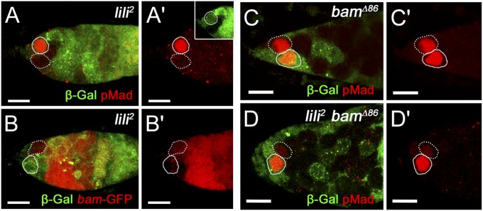
lili is required for BMP signaling in the GSC. In all images, homozygous clonal GSCs are marked by a dashed outline, whereas neighboring nonclonal GSCs are marked by a solid outline. (A–B′) Germaria containing a lili2 mutant GSC (β-gal negative; green) next to a WT GSC (β-gal positive; green) stained for pMad (A and A′) or bamP-GFP (B and B′) expression (both shown in red). The lili mutant GSC have lower pMad and higher bamP-GFP compared with neighboring WT GSCs. (C and C′) Germarium containing a bamΔ86 mutant GSC (β-gal negative; green) next to a WT GSC (β-gal positive; green) stained for anti-pMad (red). Robust pMad was present in all clonal GSCs observed (n = 38). (D) Germarium containing a clonal lili2 bamΔ86 double mutant GSC (β-gal negative; not green) next to a WT GSC (β-gal positive; green) stained for pMad (red). Loss of pMad is observed even in the absence of differentiation. (Scale bars: 10 µm.)

