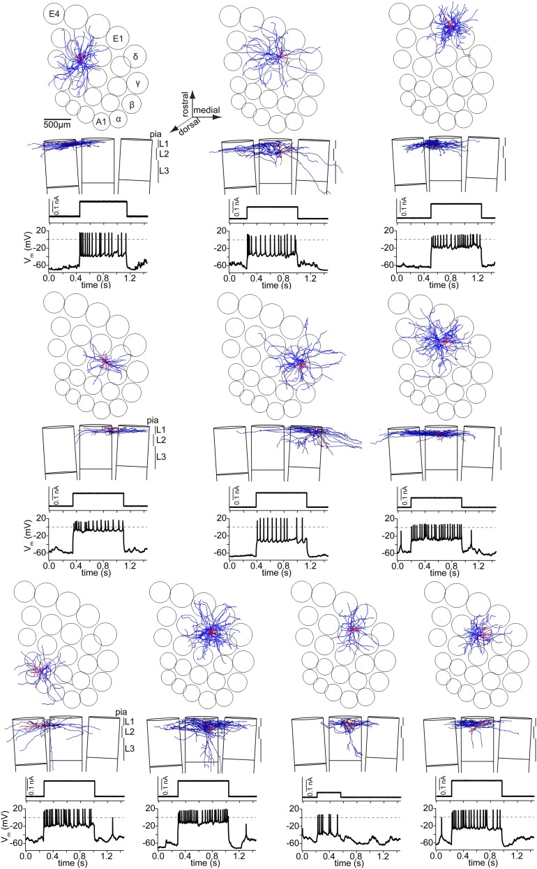
Fig. S2.
L1 IN axon morphologies. (Top) Tangential view onto vS1 showing the neuron reconstructions (axon, blue; dendrites and soma, red) at their registered location within the vS1 model. (Middle) Coronal view, showing the supragranular layers of three adjacent barrel columns in the same row and the vertical location of the neuron reconstruction. (Bottom) Response to current injection in vivo.

