Fig. 1.
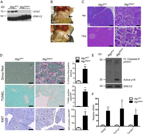
Atg7∆pan mice exhibit pancreatic degeneration, inflammation, and fibrosis. (A) IB analysis of ATG7 in pancreatic lysates of 12-wk-old mice of indicated genotypes. (B) Gross morphology of pancreata in 12-wk-old Atg7F/F and Atg7∆pan mice. (C) H&E staining of pancreatic tissue sections from 4- and 12-wk-old mice. (Scale bars, 100 μm.) (D) Histological analysis (IHC) of pancreatic sections from 12 wk old Atg7F/F and Atg7∆pan mice. Sirius Red, TUNEL, and Ki67 staining, and corresponding quantitation of positive cells per high power (200×) field (HPF). (Scale bars, 100 μm.) (E) IB analysis of caspase 8 in pancreatic lysates from 3-wk-old mice. ERK1/2: loading control. (F) qPCR analysis of α-SMA (Acta2), collagen1A(I) (Col1a1) and collagen 3A(I) (Col3a1) mRNAs in 12-wk-old mice of indicated genotypes. Values in D and F are means ± SEM n = 3–4 mice per condition. *P < 0.05, **P < 0.01, ***P < 0.001.

