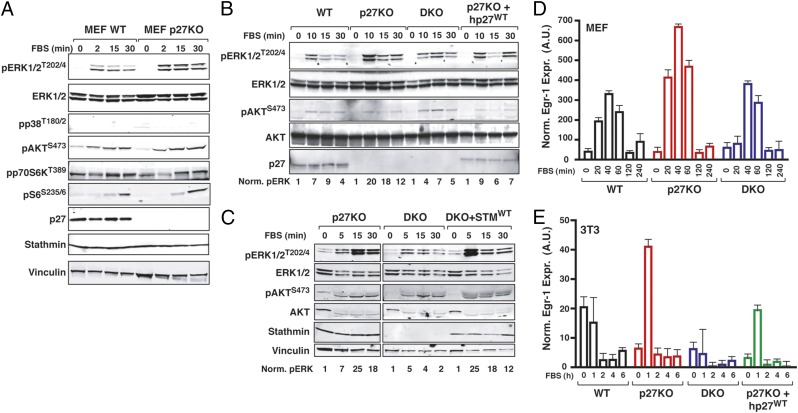
Fig. 2.
Interaction of p27/stathmin regulates MAPK pathway activation. (A) Western blot analysis of the mTOR and MAPK pathway activation in WT and p27KO MEFs that were serum-starved and then stimulated with 10% serum (FBS) for the indicated times. Western blot analysis of pERK1/2 and pAKT in 3T3 fibroblasts of the indicated genotypes and 3T3 p27KO stably reexpressing p27 (B) or 3T3 DKO stably reexpressing stathmin (C) was performed. Cells were serum-starved and then stimulated with FBS for the indicated times. Numbers at the bottom of the panels indicate the quantification of normalized (Norm.) ERK1/2 phosphorylation levels. Quantitative RT-PCR analysis of Egr-1 in MEFs (D) or 3T3 fibroblasts (E) that were serum starved and then stimulated with 10% serum (FBS) for the indicated times. A.U., arbitrary units.

