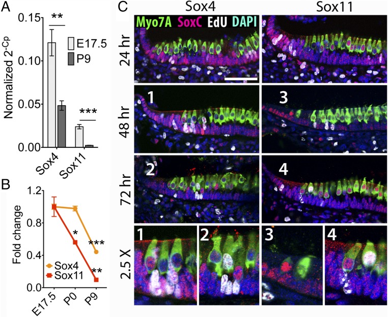
Fig. 2.
SoxC expression in the developing sensory organs of the inner ear. (A) qPCR confirms the decrease in expression of SoxC genes between E17.5 and P9. The significance of the change in expression is **P = 0.0012 for Sox4 (n = 6) and ***P = 0.0001 for Sox11 (n = 6). (B) Normalized to the RNA-sequencing data from E17.5 mice, the fold changes in the number of reads per kilobase of exon per million fragments mapped emphasize the delay in the down-regulation of Sox4 relative to Sox11. The change is significant at P0 for Sox11 (*P = 0.023, n = 3) and at P9 for both Sox4 (***P = 0.0001, n = 3) and Sox11 (**P = 0.0017, n = 3). (C) Twenty-four hours after injection, EdU labeling and antibody labeling for Sox4 and Sox11 (red) demonstrate that these proteins occur in actively proliferating supporting cells (white) at the periphery of each utricular macula. Some labeled hair cells (green) are newly formed by the criteria of EdU labeling, peripheral localization, absence of mature hair bundles, and weak immunolabeling for Myo7A. Representative SoxC-expressing hair cells are shown in the bottom images at a further enlargement of 2.5×. Nuclei are labeled blue. (Scale bar, 50 μm.)

