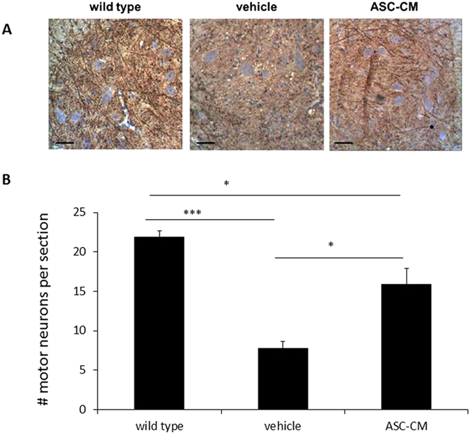
Figure 2. ASC-CM 7-day treatment of SOD1G93A mice with disease showed increase number of motor neurons in the lumbar spinal.

SOD1G93A mice were given ASC-CM or vehicle for 7 days after disease onset. Motor neurons in the spinal cord area were identified by immunoreactivity to MAP2 antibody. Representative images showed an increased number of MAP2-positive neurons in lumbar spinal cords of SOD1G93A mice treated with ASC-CM compared to vehicle. Scale bars: 100 μm. (A). MAP2 immunoreactive images covering the entire cross-sectional area from6–8 lumbar spinal regions for each mouse were quantitated. With ASC-CM treatment, a higher number of motor neurons was observed in SOD1G93A mice with disease onset (n = 5) versus vehicle (n = 5). Wild type control data were generated from 4 mice (B). Experimental group data are presented as averages (±SEM) and statistical analyses were performed using one-way ANOVA. *p < 0.05; ***p < 0.005.
