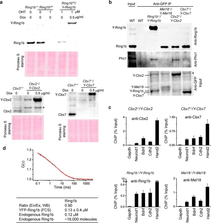
FIGURE 4.
Fusion proteins tested recapture the functions of their endogenous counterparts. a, Western blot analysis of protein levels using antibodies directed against endogenous proteins. Ponceau S staining was used for the loading control. * indicates nonspecific bands. b, IP analysis of the interaction of endogenous Ring1b and Phc1 with YFP-PRC1 fusion proteins. Extracts were precipitated by anti-GFP antibody. The precipitates were analyzed by immunoblotting using antibodies directed against Ring1b and Phc1. The input contained 5% of the extract. WT denotes PGK12.1 mES cells. * indicates nonspecific bands. c, ChIP analysis of the binding YFP-PRC1 fusion proteins to endogenous target gene promoters. The fragmented chromatins isolated from Cbx2−/−/Y-Cbx2, Cbx7−/−/Y-Cbx7, Ring1b−/−/Y-Ring1b, and Mel18−/−/Y-Mel18 mES cells were precipitated using antibodies directed against Cbx2, Cbx7, Ring1b, and Mel18, respectively. Results are means ± S.D. d, quantification of the number and the concentration of Ring1b·PRC1 complexes in mES cells. The autocorrelation curves (black dot line) were fitted with the one component model of free diffusion in three dimensions with triplet function (red line). The table shows the ratio of endogenous to YFP-tagged Ring1b protein (En/Ex) detected by Western blotting (WB), the concentration of endogenous Ring1b and YFP-Ring1b fusion, and the number of endogenous Ring1b proteins.

