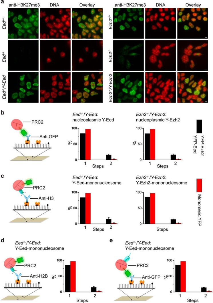
FIGURE 9.
PRC2 is a mixture of monomer and dimer and binds to mononucleosome in a 1:1 or 2:1 stoichiometry. a, immunostaining of H3K27me3 in Ezh2+/+, Eed+/+, Ezh2−/−, Eed−/−, Ezh2−/−/Y-Ezh2, and Eed−/−/Y-Eed mES cells by using antibody directed against H3K27me3 (green). DNAs were stained with Hoechst (blue). Overlay images are shown. Scale bar is 5 μm. b, nucleoplasmic YFP-Eed and YFP-Ezh2 are a mixture of monomers and dimers. The YFP-PRC2 complexes extracted from Ezh2−/−/Y-Ezh2 and Eed−/−/Y-Eed mES cells were pulled down by biotinylated anti-GFP antibody via interaction with NeutrAvidin (left). The percentage of fluorescence photobleaching steps of YFP-Eed and YFP-Ezh2 is shown as black bar (right). For a comparison, the red bar for the monomeric YFP is replicated from Fig. 1b. Results are means ± S.D. c, PRC2 binds to mononucleosome in a 1:1 or 2:1 stoichiometry. The YFP-PRC2·mononucleosome complexes from Ezh2−/−/Y-Ezh2 and Eed−/−/Y-Eed mES cells were immobilized by biotinylated antibodies directed against H3 (left). The percentage of fluorescence photobleaching steps of YFP-Eed and YFP-Ezh2 on a mononucleosome is shown as black bar (right). Results are means ± S.D. For comparison, the red bar for the monomeric YFP is replicated from (Fig. 1b). d and e, percentage of fluorescence photobleaching steps of YFP-Eed on a mononucleosome. The YFP-Eed·PRC2·mononucleosome complexes were immobilized by biotinylated antibodies directed against H2B (d) and GFP (e). For a comparison, the red bar for the monomeric YFP is replicated from Fig. 1b. Results are means ± S.D.

