FIGURE 3.
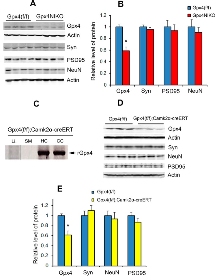
Gpx4 ablation resulted in no overt neurodegeneration in the cerebral cortex. A, Western blots showing levels of GPX4, Syn, NeuN, and PSD95 in cortex tissues from Gpx4(f/f) mice and Gpx4NIKO mice on day 6 post TAM treatment (n = 9, male and female). B, quantified levels of GPX4, Syn, NeuN, and PSD95 in cortex tissues. *, p < 0.05. C, detection of rGpx4 by PCR only in nervous tissues from TAM-treated Gpx4(f/f);Camk2α-creERT mice. The image was assembled from different parts of a gel. CC, cerebral cortex; HC, hippocampus; SM, skeletal muscle; Li, liver. D, Western blots showing levels of GPX4, Syn, NeuN, and PSD95 in cortex tissues from Gpx4(f/f) mice and Gpx4(f/f);Camk2α-creERT mice 2 weeks post-TAM treatment. E, quantified levels of GPX4, Syn, NeuN, and PSD95 in cortex tissues from Gpx4(f/f) mice and Gpx4(f/f);Camk2α-creERT mice 2 weeks post-TAM treatment (n = 3, male and female). *, p < 0.05.

