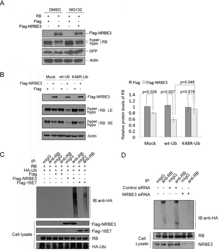
FIGURE 3.
NRBE3 promoted RB degradation through proteasome-ubiquitination pathway. A, RB and GFP plasmids were co-transfected either with FLAG-NRBE3 or FLAG vector plasmid into H1299 cells. Cells were treated either with 10 μm MG132 or dimethyl sulfoxide (DMSO) for 4 h before harvest. Equal amounts of protein of whole cell lysates were subjected to Western blotting for the indicated proteins. GFP was evaluated as a transfection efficiency control, and β-actin was evaluated as a loading control. B, H1299 cells were transfected with FLAG-NRBE3 or FLAG vector plasmid in the presence of HA-tagged wild-type ubiquitin plasmid (wt-Ub), HA-tagged K48R ubiquitin mutant plasmid (K48R-Ub), or empty vector (Mock). Equal amounts of protein from whole cell lysates were subjected to immunoblotting for evaluation of expression of FLAG-NRBE3 and RB as indicated. β-Actin was evaluated as a loading control. LE, long exposure bands; SE, short exposure bands (left panel). -Fold induction of the relative protein levels of RB is summarized from three independent experiments. Error bars represent S.E. (right panel). C, H1299 cells were co-transfected with RB, HA-Ub, and FLAG-NRBE3, FLAG-HPV16 E7, or FLAG vector plasmids, respectively. Cells were treated with 10 μm MG132 for 4 h before harvest. RB protein was immunoprecipitated from cell lysates with anti-RB antibody or mouse IgG. Proteins from the precipitates were subjected to Western blotting with anti-HA antibody (upper panel). Expression of RB, FLAG-NRBE3, FLAG-HPV16 E7, or HA-Ub was evaluated by immunoblotting with anti-RB, anti-FLAG anti-E7, or anti-HA antibody on cell lysates as indicated (lower panel). D, HCT116 cells were co-transfected with HA-Ub and NRBE3-specific siRNA or control siRNA, respectively. Cells were treated with 10 μm MG132 for 4 h before harvest. RB protein was immunoprecipitated from cell lysates with anti-RB antibody or mouse IgG. Proteins from the precipitates were subjected to Western blotting with anti-HA antibody (upper panel). NRBE3 and RB in cell lysates were evaluated by immunoblotting with anti-NRBE3 and anti-RB (lower panel). mIgG, mouse IgG; IP, immunoprecipitation; IB, immunoblot, hypo, hypophosphorylated; hyper, hyperphosphorylated.

