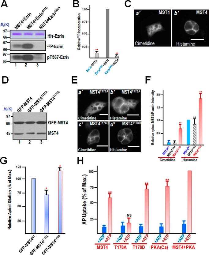
FIGURE 4.
PKA-primed phosphorylation of Ser-66 promotes the phosphorylation of Thr-567 by MST4. A, MST4 exhibits better efficiency in phosphorylation of ezrin-mimicking Ser-66 phosphorylation in vitro. Aliquots of recombinant hexahistidine-tagged ezrin, including wild-type and ezrin mutants (S66A and S66D), were incubated with MST4 as detailed under “Materials and Methods.” Top panel, Coomassie Blue-stained SDS-PAGE gel. Bottom panel, 32P incorporation from the same gel. The efficiency of MST4-elicited 32P incorporation into ezrin is highest in the ezrinS66D mutant (lane 2) and very little in the ezrinS66A mutant (lane 3). B, quantitative analyses of in vitro phosphorylation efficiency of Thr-567 using various ezrin proteins mimicking Ser-66 phosphorylation. **, p < 0.01 compared with EzrinS66D. Error bars represent the mean ± S.E. of three separate preparations. C, immunofluorescence staining of MST4 in non-secreting and secreting culture parietal cells. MST4 is primarily located to the cytoplasm, with a slight concentration at the apical membrane (a'). However, after histamine stimulation, the MST4 signal is enriched in dilated apical membrane vacuoles (b'). Scale bar = 10 μm. D, characterization of expression of GFP-MST4 proteins in gastric parietal cells. Aliquots of parietal cells were transiently transfected to express MST4 proteins (wild-type, T178A, and T178D). 24 h after transfection, cell lysates were prepared and fractionated on SDS-PAGE, followed by Western blot analyses with MST4 antibody. Note that the exogenous MST4 proteins expressed 3-fold higher compared with endogenous MST4. E, PKA-activated MST4 promotes parietal cell activation by histamine stimulation. Aliquots of parietal cells were transiently transfected to express MST4 proteins (T178A and T178D). 24 h after transfection, the parietal cells were treated with histamine (100 μm) as detailed under “Materials and Methods,” followed by fixation and immunofluorescence analyses. Note that the exogenous MST4T178D-expressing cells exhibited larger apical membrane vacuoles even before histamine stimulation (c'). Scale bars = 10 μm. F, quantitative analyses of apical localization of MST4 (WT; phospho-mimicking, T178D; non-phosphorylatable, T178A) in cimetidine- and histamine-treated parietal cells. **, p < 0.01 compared with wild-type MST4-expressing cells in secreting and non-secreting status. Error bars represent the mean ± S.E. of three separate preparations of 20 cells in each category. G, quantitative analyses of apical vacuole diameter of MST4-expressing parietal cells as a function of activation. *, p < 0.05 compared with stimulated controls. Max., maximal stimulation. Error bars represent the mean ± S.E. of three separate preparations of 50 cells in each category. H, MST4 cooperates with PKA for optimal parietal cell activation. Aliquots of gastric glands were permeabilized with SLO, and various MST4 recombinant proteins and PKA were added to the permeable cells for secretory activity measurements using the AP uptake assay, as described under “Materials and Methods.” NS, no significant difference; **, p < 0.001; *, p < 0.05 compared with stimulated controls. Error bars represent the mean ± S.E. of three separate preparations.

