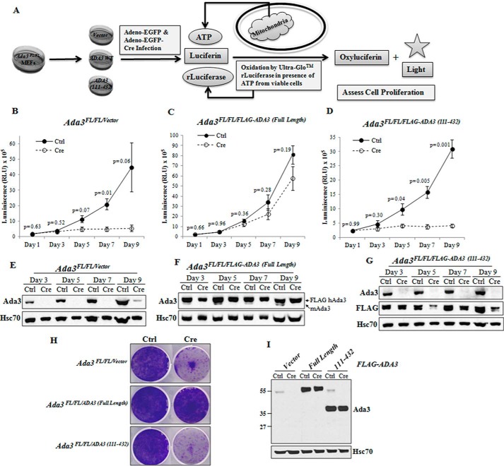
FIGURE 4.
CENP-B binding-defective ADA3 mutant fails to rescue cell proliferation arrest caused by the deletion of endogenous Ada3. A, strategy used to perform ADA3 rescue cell proliferation assays. B–D, cell viabilities of Ada3FL/FL/Vector (B), Ada3FL/FL/FLAG-ADA3(Full-Length) (C), and Ada3FL/FL/FLAG-ADA3(111–432) (D) MEFs after control adenovirus (Ctrl) or Cre adenovirus (Cre) infection obtained using a CellTiter-GLO luminescent cell viability assay as described under “Experimental Procedures.” Data shown here are mean ± S.E. (error bars) from three independent experiments performed in six replicates, and p values were computed using Student's t test. E–G, ADA3 protein levels at different time points after Cre adenovirus infection of the indicated cell lines. Note that full-length or mutant ADA3 reconstituted control cells express both mouse ADA3 and human FLAG-ADA3 proteins, whereas only human FLAG-ADA3 is seen in Cre adenovirus-infected cells. H, colony formation assay. Crystal violet staining of the indicated cells infected with control virus or Cre adenovirus grown for 9 days is shown. I, Western blotting of lysates from H showing exogenous and endogenous ADA3.

