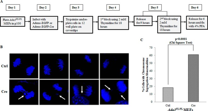
FIGURE 6.
Deletion of Ada3 causes defects in chromosome segregation. A, strategy used to assess chromosome mis-segregation in Ada3-deleted cells. B, representative confocal images of DAPI-stained anaphase chromosomes from control or Cre-infected Ada3FL/FL MEFs. Note that Ada3-deleted cells show various chromosomal segregation abnormalities, such as lagging chromosomes and anaphase bridges (indicated by white arrows). C, quantification of chromosomal segregation defects in control or Cre-infected Ada3FL/FL MEFs from B. Note that at least 50 anaphase cells each from control (Ctrl) and Cre-infected Ada3FL/FL MEFs were analyzed for quantification.

