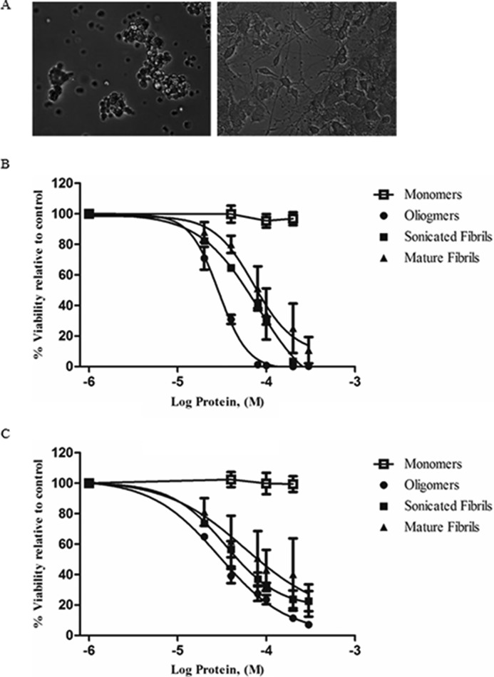
FIGURE 3.

Effects of NGF treatment on cultured PC12 cells. A, light microscopic image of undifferentiated (left) and 100 ng/ml of treated differentiated cells (right) (scale bar: 100 μm). B and C, Alamar Blue emission viability results for PC12 cell lines treated with HEWL-separated fractions (Log scale). Undifferentiated (B) and differentiated (C) PC12 cells were exposed to varying concentrations of prefibrillar, and fibrillar aggregates then assayed using the fluorescent staining compound Alamar Blue. Monomeric HEWL was also examined and displayed no decrease in viability. The error bar indicates the values of mean ± S.E. of three experiments and were calculated using GraphPad software.
