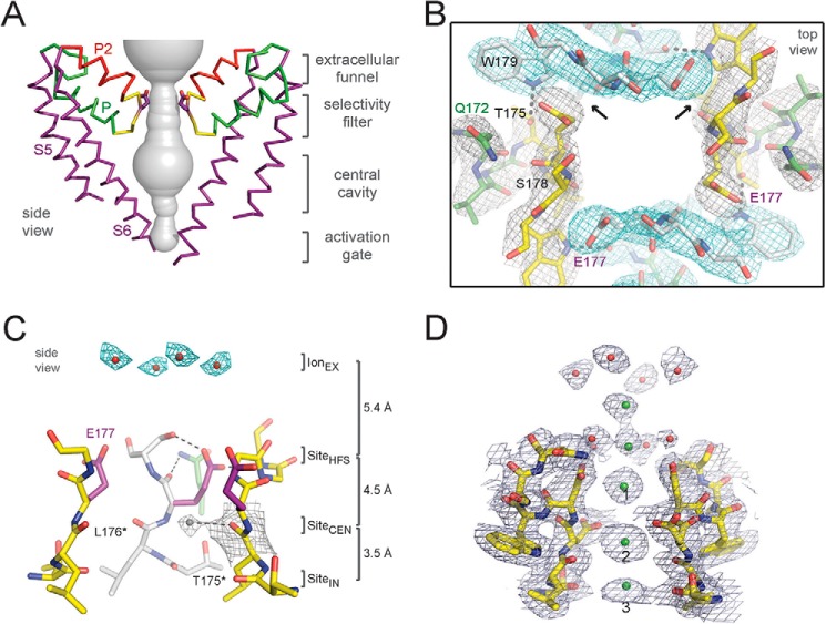
FIGURE 8.
Structure of the pore and selectivity filter of NavAb and CavAb. A, architecture of the NavAb pore. Purple, Glu177 side chains; gray, pore volume. The S5 and S6 segments and the P loop from two lateral subunits are shown (53). B, top view of the ion selectivity filter. Symmetry-related molecules are colored white and yellow; P-helix residues are colored green. Hydrogen bonds between Thr175 and Trp179 are indicated by gray dashes. Electron densities from Fo − Fc omit maps are contoured at 4.0 σ (blue and gray), and subtle differences can be appreciated (small arrows) (53). C, side view of the ion selectivity filter. Glu177 (purple) interactions with Gln172, Ser178, and the backbone of Ser180 are shown in the far subunit. Fo − Fc omit map at 4.75 σ (blue) and putative cations or water molecules (red spheres, IonEX) are shown. Electron density around Leu176 (gray; Fo − Fc omit map at 1.75 σ) and a putative water molecule are shown (gray sphere). Na+ coordination sites: SiteHFS, SiteCEN, and SiteIN (53). D, CavAb selectivity filter. Electron density at the selectivity filter of 175TLDDWSN181 is shown. The 2Fo − Fc electron density map (contoured at 2 σ) of select residues in the selectivity filter with two diagonally opposed subunits is shown in stick format, the Ca2+ ions along the ion pathway are shown in green spheres, and water molecules are shown in red spheres. Note that it is likely that adjacent calcium-binding sites are not occupied simultaneously because of electrostatic repulsion. Our model is that the site at Asp181 in the outer vestibule and the high-field-strength site at Asp177 are occupied together, the sites at Asp178 and the carbonyl sites at Leu176/Thr175 are occupied together, and the selectivity filter and vestibule oscillate between these two ion occupancies to mediate rapid conductance.

