FIGURE 3.
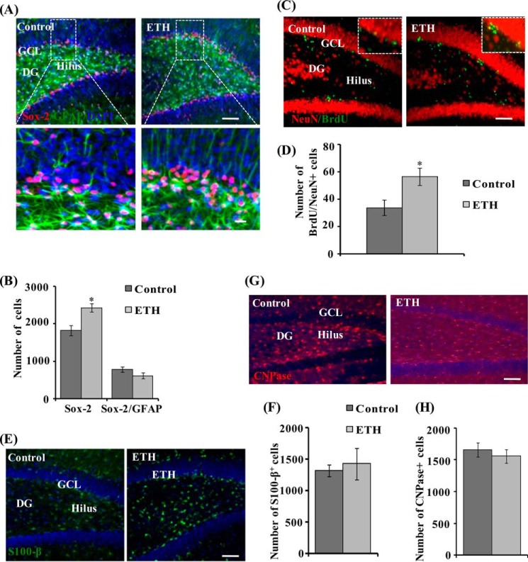
ETH enhances the NPC pool and neuronal differentiation in the hippocampus of adult rats. A, photomicrographs showing immunostaining of Sox-2+ cells (red) co-labeled with GFAP (green) and DAPI (blue) in the dentate gyrus region of the hippocampus. Most of the Sox-2/GFAP+ cells were found in the hilus and GCL regions, whereas a few Sox-2/GFAP+ cells were also present in the molecular layer (ML) of the DG. Arrows, Sox-2/GFAP+-co-labeled cells. Inset, higher magnification of Sox-2+ cells and Sox-2/GFAP-co-labeled cells. Scale bar, 100 μm (A) and 10 μm (inset). B, quantitative analysis of the number of Sox-2+ cells and Sox-2/GFAP+-co-labeled cells. Values are expressed as mean ± S.E. (error bars) (n = 6 rats/group). *, p < 0.05 versus control. C, double immunofluorescence images of matured neurons co-labeled with NeuN (red; mature neuronal nuclei marker) and BrdU (green) in the DG region of the hippocampus. Inset, higher magnification images. Scale bar, 100 μm. D, quantitative analysis of NeuN/BrdU-co-labeled cells in the hippocampus. E, images showing immunostaining of the astrocyte-specific marker (S100-β) in the hippocampus. Scale bar, 100 μm. F, quantitative analysis of S100-β+ cells in the hippocampus. G, images showing immunostaining of the oligodendrocyte-specific marker (CNPase) in the hippocampus. Scale bar, 100 μm. H, quantitative analysis of CNPase+ cells in the hippocampus.

