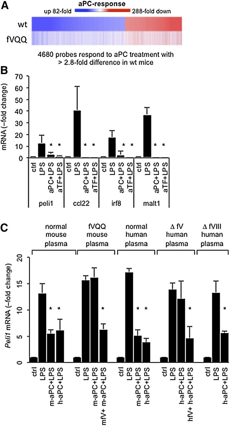
Figure 3.
aPC resistance of fV Leiden suppresses regulation of inflammatory gene expression by aPC. (A) Heat map depicts the regulation of 4680 genes that are responsive to aPC therapy in spleen dendritic cells collected by fluorescence-activated cell sorting 16 hours after LPS (LD50) challenge from a wild-type (wt) mouse as compared to a homozygous fV Leiden mouse (fVQQ). Equal amounts of total RNA were pooled from 5 individual animals per genotype and analyzed by hybridization to gene arrays. Colors indicate the response (fold change up or down) compared to LPS-challenged mice not receiving aPC. Individual genes are sorted according to their aPC response in wild-type mice. (B) Peli1, Ccl22, Irf8, and Malt1 mRNA levels were measured by quantitative real-time PCR analysis in RAW cells cultured with 10% normal mouse plasma (ctrl) and cells treated for 3 hours with 100 ng/mL of LPS, 100 ng/mL of LPS plus 100 ng/mL of mouse aPC (aPC+LPS), or 100 ng/mL of LPS plus 5 μg/mL of anti-mouse TF antibodies (aTF+LPS). Data represent the average ± standard deviation from ≥3 independent experiments with triplicate measurements of each sample. RNA levels in control cultures were arbitrarily set to “1.” *P < .05 by pairwise comparison with LPS-induced levels (Student 2-tailed t test). (C) Pellino-1 (Peli1) mRNA, measured 3 hours after LPS (100 ng/mL) addition in response to human aPC (h-aPC) or mouse aPC (m-aPC) in the presence of (10% vol/vol) pooled normal mouse or human plasma, or pooled normal human plasma depleted of fV (Δ fV) or fVIII (Δ fVIII). Recombinant mouse fV and plasma-derived human fV were added at 100 ng/mL. *P < .05 by pairwise comparison with LPS-induced levels (Student 2-tailed t test).

