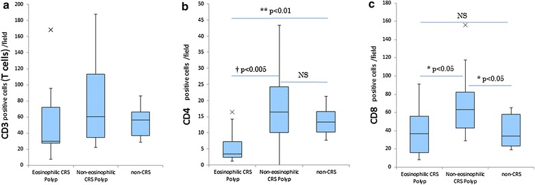
Fig. 4.

The number of inflammatory cells in mucosal specimens. The number of CD3 positive cells (T cells) (a), CD4 positive cells (b) and CD8 positive cells (c) per field in the ECRS and non-ECRS polyps, as well as the non-CRS controls. Data in box-and-whisker plots represent the median, lower and upper quartile and the minimum to maximum value. ×, outliers (†p < 0.005, **p < 0.01, *p < 0.05)
