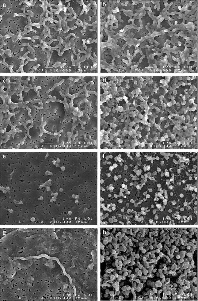
Fig. 3.

Micrographs of C. jejuni NCTC 11168 and C. jejuni Bf in MAC (a, b, respectively), and AC (c, d, respectively) after 12 h of incubation, and C. jejuni NCTC 11168 and C. jejuni Bf in MAC (e, f, respectively), and AC (g, h, respectively) after 24 h of incubation using SEM on polycarbonate membranes. Magnification: ×10,000
