Fig. 2.

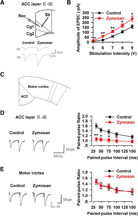
Enhancement of synaptic transmission in the ACC of animal model of IBS. a The location of stimulation and recording (top), and representative synaptic input–output curves in the ACC slices from control and zymosan-injected mice (bottom). b The amplitude of EPSCs was obviously enhanced in the ACC slice of mice injected with zymosan (n = 11/6 mice) as compared with that of control mice (n = 10/6 mice). c The location of the ACC and motor cortex in coronary slice. d Paired-pulse ratio (the ratio of EPSC2/EPSC1) was recorded at intervals of 35, 50, 75, 100, and 150 ms from control (n = 9/6 mice) and zymosan-injected mice (n = 9/6 mice). PPF was markedly reduced in the ACC of mice injected with zymosan at intervals of 35, 50, 75, and 100 ms. e PPF in motor cortex neurons had no difference in control (n = 9/6 mice) and zymosan-injected mice (n = 11/6 mice). * P < 0.05, ** P < 0.01 vs. control
