Fig. 3.

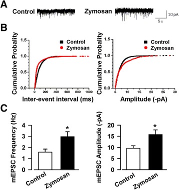
Enhanced mEPSCs in the ACC of animal model of IBS. a Representative mEPSCs recorded in pyramidal neurons at a holding potential of −70 mV from control and zymosan-injected mice. b Cumulative inter-event interval (left) and amplitude (right) histograms of mEPSCs recorded in slices of control (n = 10/6 mice) and zymosan-injected mice (n = 11/6 mice). c Summary plots of mEPSC data. The frequency (left) and amplitude (right) of mEPSCs were significantly enhanced in the ACC slices of mice injected with zymosan. * P < 0.05 vs. control
