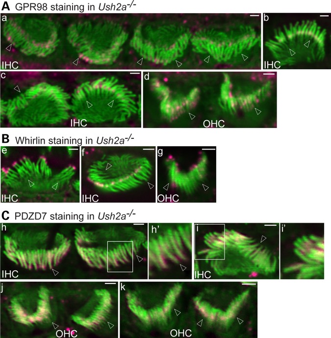
Figure 4.
Usherin plays differential roles in localization of GPR98, whirlin and PDZD7 at the cochlear ALC. (A) GPR98 (magenta) distribution at the ALC is altered in most IHCs and OHCs of P4 Ush2a−/− mice. GPR98 punctate signals are localized predominantly at both the tip and base of Ush2a−/− IHC stereocilia (a–c). (a) Back view of four Ush2a−/− IHC stereociliary bundles showing abnormal GPR98 distribution; (b) front view of an Ush2a−/− IHC bundle showing normal GPR98 distribution; (c) side view of an Ush2a−/− IHC bundle with abnormal GPR98 signal (left) and front view of another Ush2a−/− IHC bundle with near-normal GPR98 signal (right); (d) GPR98 punctate signals are randomly distributed along Ush2a−/− OHC stereocilia. (B) Whirlin (magenta) is completely absent at the stereociliary base but detectable at the stereociliary tip in both P4 Ush2a−/− IHCs and OHCs. (e and f) Front and back views of Ush2a−/− IHC bundles, respectively; (g) back view of an Ush2a−/− OHC bundle. (C) PDZD7 (magenta) distribution in Ush2a−/− IHC (h and i) and OHC (j and k) stereociliary bundles is similar to that in Adgrv1−/− IHC and OHC bundles at P4. Insets (h′ and i′) are amplified views and shown in original images for context. Images (h), (j) and (k): back view; (i): side view. Arrows point to stereociliary bases. Green signals are from phalloidin. Magenta signals outside stereociliary bundles are non-specific. Scale bars, 1 μm.

