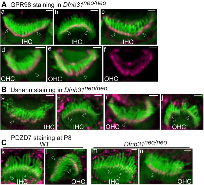
Figure 5.
Minor mislocalization of GPR98 and normal localization of usherin and PDZD7 in Dfnb31neo/neo cochlear hair cells. (A) GPR98 (magenta) is localized normally at stereociliary bases of most P4 Dfnb31neo/neo IHC (a, b, back and front views, respectively) and OHC (d) bundles, while GPR98 is partially mislocalized to stereociliary tips in a small number of Dfnb31neo/neo IHCs (c) and OHCs (e). (f) Single magenta channel image of (e). (B) Usherin signals (magenta) remain at stereociliary bases of P4 Dfnb31neo/neo cochlear hair cells. (g and h) Back and front views of Dfnb31neo/neo IHC bundles, respectively; (i and j) back and front views of Dfnb31neo/neo OHC bundles, respectively. (C) PDZD7 (magenta) localization appears normal in P8 Dfnb31neo/neo IHC (m) and OHC (n) relative to age-matched wild-type (WT) IHC (k) and OHC (l). Arrows point to stereociliary bases. Green signals are from phalloidin labeling. Magenta signals outside stereociliary bundles are non-specific. Scale bars, 1 μm.

