Figure 1.
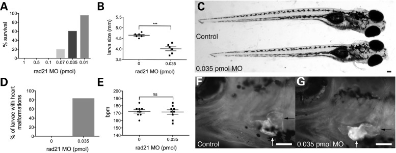
Partial depletion of Rad21 produces heart defects in zebrafish larvae that otherwise appear normal. Zebrafish embryos were injected at the one-cell stage with 1 nl phenol red dye, or with various quantities of Rad21 ATG MO, then raised at 25°C. (A) Percentage of Rad21-depleted larvae surviving to swim and eat normally at 12 dpf (n > 20 for each condition). (B) Larval size at 12 dpf. Rad21-depleted larvae were significantly smaller than control embryos (P < 0.05, unpaired t-test). n = 6 for each condition, error bars are ±SEM. (C) Rad21-depleted larva (below) and control (top) at 12 dpf. Dorsal view, anterior to the right. Apart from their slightly smaller size, Rad21-depleted larvae appeared externally normal. Scale bar = 100 µm. (D) Percentage of larvae displaying a heart defect and reduced circulation at 12 dpf. Eighty-three percent of Rad21-depleted larvae (n = 10/12) displayed this phenotype. Heart defects were never observed in control larvae (n = 20/20). (E) Heartbeats per minute (bpm) at 12 dpf. Heartbeat did not differ between Rad21-depleted larvae and controls (P > 0.05, unpaired t-test). n = 9 for each condition, error bars are ±SEM. (F and G) Heart morphology at 12 dpf, visualized using a double-transgenic Tg(gata1:dsred), Tg(β-actin:GFP) zebrafish line. Black arrows indicate the blood-filled atrium, white arrows indicate the ventricle. Ventral views, anterior to the left. Scale bars are 100 µm. In control larvae (F), the orientation of the atrium and ventricle were at right angles, whereas in Rad21-depleted larvae (G), the orientation of the atrium and ventricle were more aligned (see Supplementary Material, Video S1).

