Figure 4.
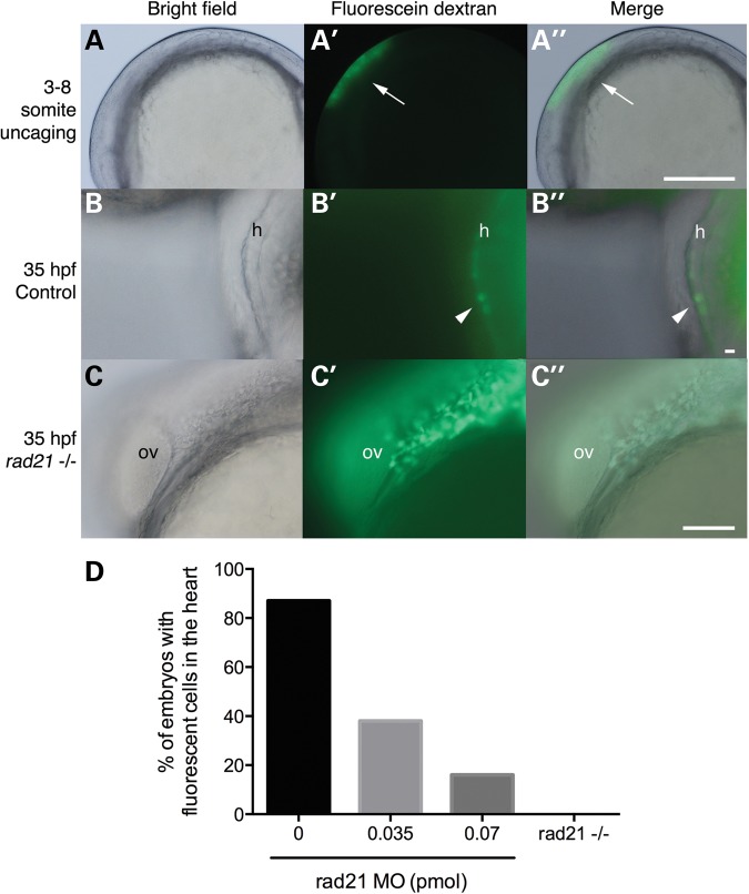
Cardiac neural crest cells fail to contribute to the heart in Rad21-depleted embryos. Wild-type, Rad21-depleted or rad21nz171 homozygous mutant zebrafish embryos were injected at the one-cell stage with caged fluorescein dextran. (A–A″) Uncaging of fluorescein dextran in the pre-migratory neural crest at three- to eight-somite stages (10.5–11.5 hpf). (A) Brightfield image of an embryo at the six-somite stage. (A′) Fluorescein dextran was uncaged in the region along the neural tube cranial to somite 1 by exposure to 360 nm UV light. (A″) A merged view shows the position of the uncaged region. Scale bar is 250 µm. (B–B″) In control embryos, fluorescein-labeled cardiac neural crest cells were present in the myocardium by 35 hpf. (B) Brightfield image of the heart (h) of a control embryo. (B′) Darkfield image showing fluorescent cardiac neural crest cells in the heart. (B″) Merged images reveal that the fluorescent cells are part of the myocardium. Scale bar is 10 µm. (C–C″) In rad21nz171 mutants, fluorescein-labeled neural crest cells never reached the heart, but instead, accumulated posterior to the otic vesicle by 35 hpf. (C) Brightfield image of the otic vesicle (ov) and the adjacent posterior region in a rad21 mutant embryo. (C′) Darkfield image showing fluorescent cells accumulating posterior to the otic vesicle. (C″) Merge of (C) and (C′). Scale bar is 100 µm. (D) Percentage of embryos with fluorescent cells in the heart. Rad21-depleted embryos (0.035 pmol MO, n = 16; 0.07 pmol MO, n = 7) displayed fluorescent cells in the heart less frequently than control embryos (n = 23). Fluorescent cells were never observed in the hearts of rad21nz171 mutants (n = 5).

