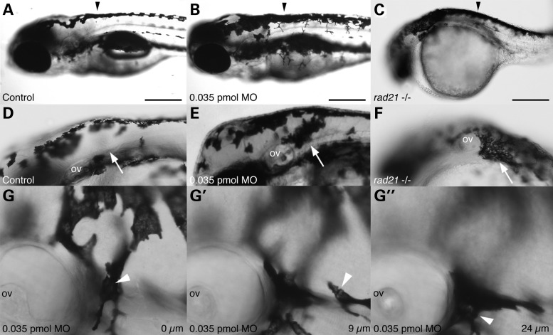
Figure 5.
Rad21 depletion causes mislocalisation of melanocytes. (A–C) Neural tube pigmentation at 3 dpf. In Rad21-depleted (0.035 pmol MO) embryos (B), we observed increased pigmentation along the neural tube compared with controls (A) (black arrowheads). Abnormal pigmentation was observed in all rad21nz171 mutant embryos (C). (D–F) Ectopic melanocytes were present behind the otic vesicle (arrows) in 15 out of 20 Rad21-depleted embryos (0.035 pmol MO) (E), and 20 out of 20 rad21nz171 mutants (F) at 3 dpf. (G–G″) Images of the ectopic melanocyte clusters of Rad21-depleted embryos, taken in different focal planes, revealed that ectopic melanocytes were present far beneath the epidermis. (G) Level of the first layer of skin (0 µm), showing one melanocyte in focus (arrowhead). (G′) Nine micrometers deeper, another melanocyte can be seen in focus (arrowhead). (G″) Twenty-four micrometers deeper, further melanocytes can be seen (arrowhead). At this developmental stage, the epidermis is <4 µm thick. The average depth of the ectopic melanocytes was 42.75 µm (n = 4). Pigmentation was not observed behind the otic vesicles of 20 out of 20 control embryos laid in the same clutch. Anterior is to the left and dorsal is to the top in all panels. Scale bars are 250 µm.

