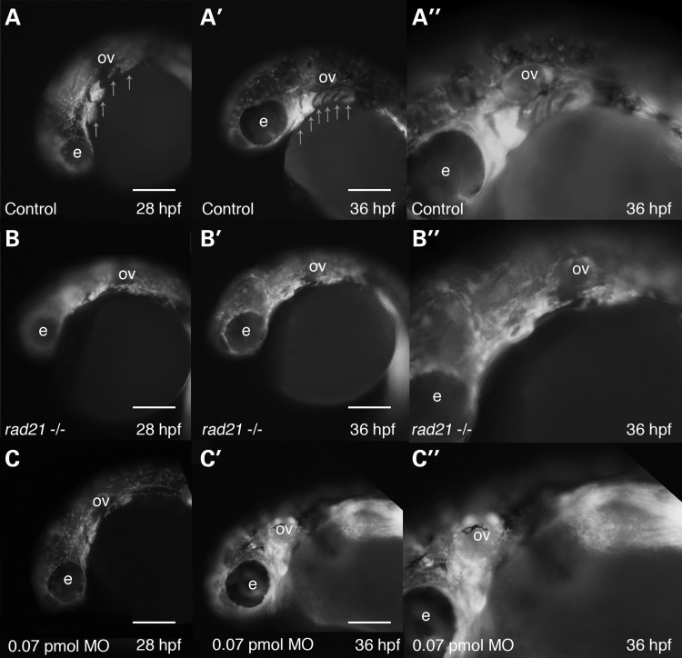
Figure 6.
Rad21 deficiency causes defects in cranial neural crest condensation into pharyngeal arch structures. Neural crest from wild-type, Rad21-depleted or rad21nz171 homozygous mutant zebrafish embryos were visualized using Tg(sox10:GFP) zebrafish. Scale bars are 250 µm. (A) In 28 hpf wild-type zebrafish embryos, neural crest cells condense and form distinctive structures representing the developing pharyngeal arches (arrows). (A′) By 36 hpf, six pharyngeal arches are clearly visible (arrows). (A″) Higher magnification of (A′) showing the pharyngeal arches in more detail. (B–B″) sox10:GFP-positive cells in rad21nz171homozygotes on the Tg(sox10:GFP) background. (B) At 28 hpf, sox10:GFP-positive neural crest cells had migrated to the anticipated location cranio-ventral to the otic vesicle (ov), but failed to correctly condense to form the characteristic arch structure. (B′) At 36 hpf, the pharyngeal arch structures remained unformed in rad21nz171 mutants. (B″) Higher magnification of (B′) showing disorganization of sox10:GFP-positive cells in the pharyngeal region. (C–C″) sox10:GFP-positive cells in Tg(sox10:GFP) embryos depleted of Rad21 using 0.07 pmol Rad21 MO. (C) Rad21-depleted embryos displayed disorganization of the pharyngeal arches at 28 hpf (18/18) as marked by sox10:GFP-positive cells. In 7 out of 18 instances, the defects recapitulated the mutant phenotype. (C′) At 36 hpf, the pharyngeal arch structures remained unformed or disorganized in Rad21-depleted embryos. (C″) Higher magnification of (C′) showing disorganization of sox10:GFP-positive cells in the pharyngeal region. See also Supplementary Material, Video S4.

