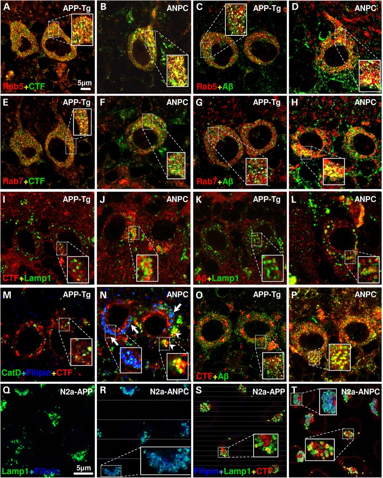
Figure 5.
Effect of EL cholesterol accumulation on intracellular localization of Aβ-related peptides in vivo and in vitro. (A–L) Representative confocal images of cerebellar Purkinje neurons in APP-Tg and ANPC mice showing localization of immunoreactive APP-CTFs (clone Y188 or C1/6.1) and Aβ peptides (clone 4G8) in Rab5-positive early-endosomes (A–D), Rab7-positive late-endosomes (E–H) and Lamp1-positive lysosomes (I–L). (M and N) Triple labeling of Purkinje neurons in APP-Tg (M) and ANPC (N) mice showing that only a subset of CatD-positive vesicles that are free of filipin-positive unesterified cholesterol contain APP-CTFs in ANPC (N, arrowheads). Arrows indicate localization of unesterified cholesterol in CatD-positive vesicle. (O and P) Representative confocal images of Purkinje neurons showing co-localization of Aβ peptides with APP-CTFs in APP-Tg (O) and ANPC (P) mice. The immunoreactive profiles of various markers were verified in three mice/genotype. (Q–T) Confocal images of N2a-APP and N2a-ANPC cells showing localization of filipin in lamp1-positive lysosomes (Q and R) and that of APP-CTFs in a subset of Lamp1-positive lysosomes that do not contain unesterified cholesterol (S and T). The immunoreactive profiles of various markers in cultured cells were verified in three independent experiments. Identity of primary antibodies is indicated by respective font colors.

