Figure 6.
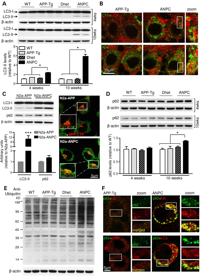
EL cholesterol impairs autophagic clearance in vivo and in vitro. (A) Immunoblots and histograms of LC3-II (∼15 kDa) in the cerebellum of 4- and 10-week-old WT, APP-Tg, Dhet and ANPC mice (n = 6 mice/genotype/age). (B) Representative double immunostaining confocal images showing colocalization of LC3-labelled puncta with APP-CTFs and Aβ. Note the increased LC3 staining intensity in Purkinje cells of ANPC versus APP-Tg mice. The profile of immunostaining was verified in three mice/genotype. (C) Quantitative analysis of LC3-II and p62 (∼62 kDa) levels along with representative confocal photomicrographs showing APP-CTFs in a subset of LC3-positive autophagic vesicles in N2a-APP/ANPC cells (n = 3). (D) Immunoblot quantitation of p62 levels in the cerebellum of 4- and 10-week-old WT, APP-Tg, Dhet and ANPC mice (n = 6 mice/genotype/age). (E) Representative immunoblot of ubiquitinated proteins in the cerebellum of 10-week-old WT, APP-Tg, Dhet and ANPC mice (n = 4 mice/genotype). (F) Representative confocal images showing co-localization of p62 with ubiquitin (upper panel) and LC3 (lower panel) in the cerebellar Purkinje neurons of ANPC and APP-Tg mice. The profile of immunostaining was verified in three mice/genotype. Identity of primary antibodies used is indicated by the respective font colors. All blots were re-probed with anti-β-actin. Data represent means ± SEM from 4 to 6 mice/genotype/age. *P < 0.05; ***P < 0.001.

