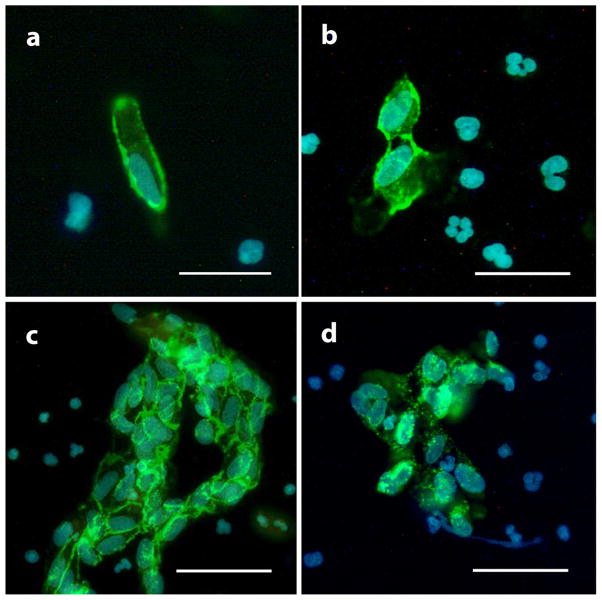
Fig. 2.
Endothelial cells identification by immunocytochemistry. ECs were identified by big size (≥20 μM), endothelial-like oval or kidney-shaped nucleus (a) and positive CD31 membrane staining (a, b and c) or vWF cytosol staining (d). Single cell (a), small aggregates of two to four cells (b) and bigger sheets of five or more cells (c) can be seen under fluorescent microscopy. Scale bar is 20 μm in (a) and (b) or 40 μm in (c) and (d).

