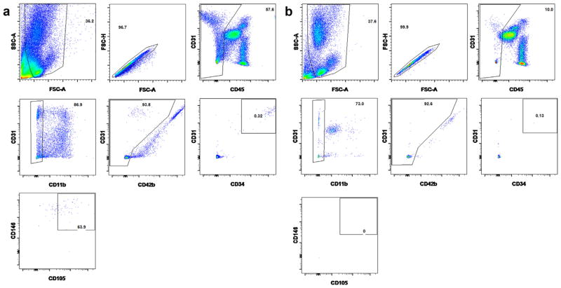
Fig. 4.
Representative figures of staining and gating strategy for live ECs on FACS. After gating on the single cells, the leukocytes (CD45+), macrophages (CD11b+) and platelets (CD42b+) were eliminated before four endothelial markers (CD31+CD34+CD105+CD146+) were used to identify and sort out the ECs. (a) and (b) Representative figures from one patient’s wire sample and peripheral blood.

