
Figure 5. RGS10 suppression alters morphology in lysophosphatidic acid-stimulated ovarian cancer cells.
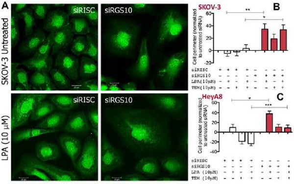
(A) SKOV-3 cells plated on glass coverslips were transfected with either siRISC or siRGS10 and treated with lysophosphatidic acid (10μM) for 30 min prior to preparation for confocal microscopy using the Whole Cell Stain Solution (ThermoFisher Scientific). (B) SKOV-3 or (C) HeyA8 cells were transfected with either siRISC or siRGS10 prior to the automated computer assessment of cell perimeter. Cells were treated with lysophosphatidic acid (LPA, 10 μM, 30 min) or temsirolimus (10 μM, ~16 h) where indicated. Cells were prepared for immunofluorescence using the Whole Cell Stain Solution and automatically scanned with high-throughput software to calculate cell perimeter. Data shows the average from 62–146 different fields per condition, which measures thousands of cells, from a series of replicate experiments and is normalized to each untreated condition (i.e. siRISC-white bars or siRGS10-shaded bars). ***p<0.001, **p<0.01 and *p<0.05 where indicated.