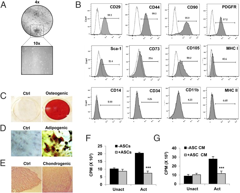
FIGURE 1.
Culture-expanded murine ASCs exhibit multilineage differentiation and immunosuppressive potential. ASCs were isolated from s.c. adipose tissue of DBA/1J mice and culture expanded as described in Materials and Methods. (A) Cells of passage 2 were analyzed for their clonogenic potential by CFU-F assay. (B) Surface phenotyping for the expression of mesenchymal and hematopoietic markers by flow cytometry. Numbers indicate percentage expression of the marker with respect to their isotype controls. (C–E) Osteogenic, adipogenic, and chondrogenic differentiation of ASCs was induced in respective differentiation media. Matrix mineralization by differentiated osteoblasts was assessed by Alizarin red S staining. Adipocytes were characterized by Oil Red O staining. Chondrogenic differentiation shows cells in lacunae in H&E-stained sections of micromass pellet cultures (original magnification ×10). (F) Suppression of activated CD4+ T cell proliferation by ASCs or (G) ASC-CM was assessed by coculturing splenic CD4+ T cells (2.5 × 10⁵ cells/ml) with ASCs (2.5 × 10³ cells/ml) or ASC-CM (50% of culture volume) under activated or unactivated conditions for 72 h. T cell activation Dynabeads were used for activating T cells, and proliferation was measured by a [3H]thymidine incorporation assay. Bar graphs are expressed as mean ± SEM of three to five replicates per group. ***p ≤ 0.001. (A)–(E) are representative of more than five batches of ASCs. (F) and (G) are representative of two independent experiments.

