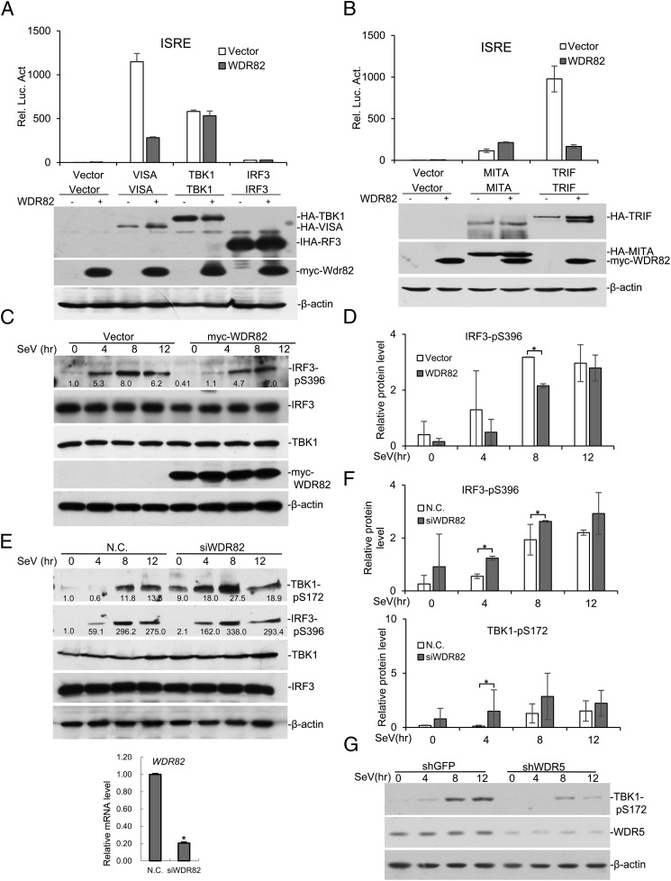
FIGURE 2.
WDR82 inhibits the phosphorylation of key signaling molecules. (A) VISA, TBK1, and IRP3 were transfected into HEK293 cells with or without WDR82. ISRE luciferase activity was assayed to measure pathway activation. (B) MITA/STING and TRIF were transfected into HEK293 with or without WDR82. Then ISRE luciferase activity was measured. (C and D) HEK293 cells were transfected with WDR82 and treated with SeV for indicated times. TBK1-pS172, IRF3-pS396, IRF3, Myc-WDR82, and β-actin were measured with Western blotting in the lysates. Quantification of Western blotting bands from three experiments are shown in (D). (E and F) HEK293 cells transfected with WDR82 siRNA were infected with SeV and the efficiency of WDR82 knockdown was confirmed by RT-PCR experiments. Western blots and protein quantification were carried out as in (C) and (D). (G) WDR5 was knocked down by shRNA and experiments were performed as in (C). Lysates were immunoblotted with anti–TBK1-pS172, anti-WDR5, and anti–β-actin. *p < 0.05.

