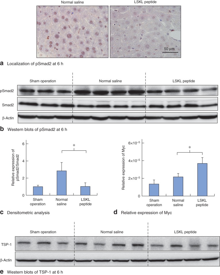
Fig. 2.

LSKL (leucine–serine–lysine–leucine) peptide successfully inhibits transforming growth factor (TGF) β–Smad signal activation induced by partial hepatectomy. a Assessment of phosphorylated Smad2 (pSmad2) nuclear localization. Immunohistochemical staining for pSmad2 at 6 h in mouse liver from normal saline and LSKL peptide groups. b Effects of LSKL peptide on pSmad2 expression in the regenerating liver at 6 h. Western blot analysis of pSmad2 in mouse liver from sham-operated, normal saline and LSKL peptide groups. β-Actin served as a loading control. c Densitometric analysis for western blots of pSmad2 in mouse liver for the three groups. d Real-time PCR analysis of relative expression of Myc mRNA in mouse liver for the three groups. c,d Values are mean(s.d.). *P < 0·010 (Student's t test). e Western blotting of thrombospondin (TSP) 1 protein at 6 h after hepatectomy. Expression of TSP-1 was similar in normal saline and LSKL peptide groups
