
Figure 3.
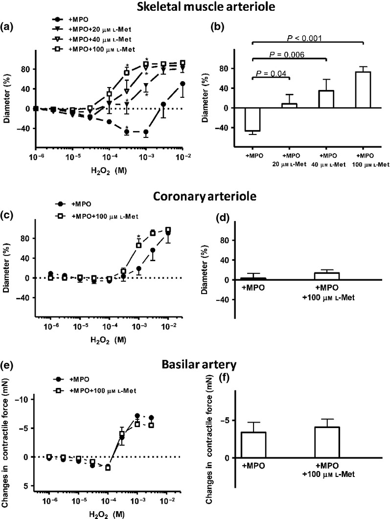
Effects of L-Met on the MPO-mediated vascular effects in different arteriolar beds. Increasing concentrations of L-Met (20, 40 or 100 μm) inhibited the MPO-mediated vasoconstriction in the SMAs in concentration-dependent manner (Id: 175 ± 24 μm, n = 6 arterioles from four different animals, with 20 μm L-Met, (closed triangles); id: 143 ± 13 μm, n = 4 arterioles from four different animals, with 40 μm L-methionine, (open triangles), id: 115 ± 19 μm, n = 5 arterioles from five different animals, with 100 μm L-Met (open squares). MPO and 20 μm L-methionine evoked significant vasoconstriction at 10 μm H2O2 compared to the baseline; panel a). The effects of MPO alone and in combination with increasing L-Met concentrations in the presence of 300 μm H2O2 (control) on the vascular diameter in the SMAs (panel b). In the CAs, L-Met (100 μm; open squares) inhibited the MPO-evoked vasoconstriction only at a higher concentration of H2O2 (id: 73 ± 10 μm, n = 4 arterioles from four different animals). Asterisks denote significant differences from MPO (panel c). The effects of MPO alone and in combination with 100 μm L-Met in the presence of 300 μm H2O2 (control) on the vascular diameter in the CAs (panel d). L-Met (100 μm; open squares) did not significantly influence the MPO-evoked changes in the isometric force in the BAs compared to the control (n = 6 arterioles from three different animals, panel e), but comparing to the zero line, MPO together with L-met caused significant vasoconstriction at 30 μm and 100 μm H2O2 (P ≤ 0.05). The effects of MPO alone and in combination with 100 μm L-Met in the presence of 300 μm H2O2 (control) on the vascular diameter in the BAs (panel f).