Figure 6.
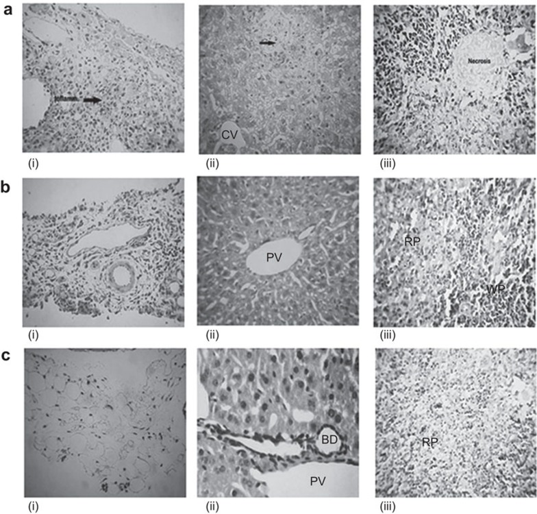
Comparative histology of different tissues from mice from the control and immunized groups. Mice from different groups were sacrificed and tissues were fixed in formalin for histopathological analysis. (a) Control mice showing (i) necrosis of villi and a severe degree of inflammation of the serosal layer of the intestine; (ii) necrosis in the hepatic parenchyma; and (iii) necrosis of the lymphoid cells in the spleen with loss of cellular outlines. (b) Mice immunized with rGroEL antigen showing (i) acute inflammatory cell infiltration in the serosal layer; (ii) normal portal triad in the liver; and (iii) improved red and white pulp areas in the splenic parenchyma compared to the control. (c) Mice to which rGroEL and rIL-22 co-administered showing (i) no inflammation of the serosal layer in the intestine; (ii) normal liver portal triad; and (iii) normal red pulp zone in the splenic tissue with sinusoids containing red blood cells (magnification, ×400). CV, central vein; PT, portal vein; RP, red pulp; WP, white pulp.

