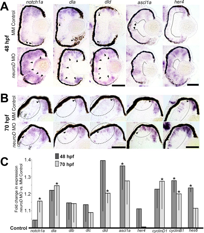
Figure 2.
NeuroD negatively regulates Notch signaling molecules and downstream targets of the Notch pathway. (A, B) In situ hybridizations showing the normal retinal expression of neurod compared to notch1a, dla, dld, ascl1a, and her4 at 48 (A) and 70 (B) hpf in control embryos and following NeuroD knockdown. Arrowheads indicate zones of expression; circled regions highlight expression differences at 70 hpf in and around the developing ONL and INL adjacent to the dorsal CMZ. Scale bars: 50 μm. (C) Quantitative RT-PCR data indicating the fold differences in expression between control embryos (black horizontal line) and embryos following NeuroD knockdown at 48 and 70 hpf. *P < 0.05, Student's t-test. Error bars: standard deviation.

