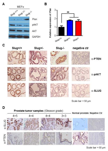
Figure 4. PTEN expression is regulated by endogenous SLUG in vivo.
(A) Western-blot analysis of AKT and PTEN expression in MEFs. Total protein was extracted from Slug+/+, Slug+/−, and Slug−/− MEFs (passage 1), and analyzed by Western-blot analysis using anti-pAKT, anti-PTEN, anti-AKT and anti-GAPDH (loading control) antibodies, respectively.
(B) qPCR analysis of PTEN transcripts in MEFs. Total RNA was extracted from the indicated MEFs and synthesized into cDNAs. Using specific primers, qPCR was used to analyze PTEN transcripts. ** p < 0.01.
(C) Expression of PTEN in prostate tissue in Slug-deficient mice. Expression of PTEN and SLUG in prostate tissues of Slug+/+, Slug+/−, and Slug−/− mice (2-month-old) was determined by immunohistochemistry using anti-PTEN, anti-pAKT, anti-SLUG antibodies, and counterstained with hematoxylin. Scale bar = 50 μm
(D) PTEN loss is related with endogenous SLUG expression in human prostate cancer tissue samples. Expression of SLUG and PTEN was detected in prostate cancer tissue samples by immunohistochemistry using anti-SLUG and anti-PTEN antibodies, and counterstained with hematoxylin. Scale bar = 50 μm.

