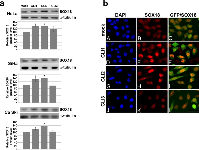
Fig 2. The effect of GLI’s overexpression on SOX18 protein level in cervical carcinoma cell lines.
a) The effect of GLI’s overexpression on SOX18 protein leveldetected by Western blot. One representative blot was presented, and quantification of protein level was presented as histogram chart.α-tubuline was used as a loading control. The relative SOX18 protein level in HeLa, SiHa and Ca Ski cells upon transfection with GLI1-3 was calculated as a percentage of SOX18 level in mock transfected cells which was set as 100%. Data of three independent experiments are presented at histograms as the means ± SEM. Values of p≤0.05 are marked by *.b)The effect of GLI’s overexpression on SOX18 protein leveldetected by immunocytochemistry. Cells were cotransfected with EGFP-C1 (that was used as a marker of transfected cells) and eiherpcDNA-mock transfection (A-C), GLI1(D-F), GLI2 (G-I), or GLI3(J-L). Boxed regions in A-L are enlarged in the same figures. Cell nuclei were counterstained with DAPI (A, D, G and J). Scale bars: 50 μm.

