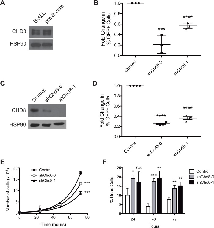
Fig 5. CHD8 depletion is detrimental in multiple B cell malignancies and normal pre-B cells.
(A) Western blot showing CHD8 expression in B-ALL cells and untransformed pre-B cells. (B) Graph showing results of growth competition assay with pre-B cells transduced with the indicated constructs. Shown are averages ± SD. Fold changes were calculated from day 2 to day 10 after retroviral infection and normalized to an empty vector control. (C) Western blot showing decreased CHD8 expression following transduction of Eμ-myc Arf -/- cells with the indicated shChd8 constructs. (D) Graph showing results of in vitro growth competition assays with Eμ-myc cells transduced with the indicated constructs. Shown are averages ± SEM of four independent experiments. Fold changes were calculated from day 2 to day 10 after retroviral infection and normalized to an empty vector control. (E) Growth curves of Eμ-myc cells transduced with the indicated constructs. Shown are averages ± SD. (F) Bar graph showing percentages of dead cells following Chd8 knockdown in Eμ-myc cells. Shown are averages ± SD. *P < 0.05, **P < 0.01, ***P < 0.001, ****P < 0.0001

