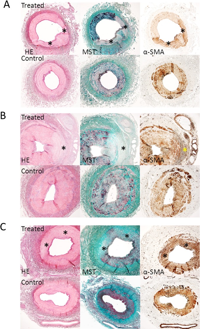
Fig 1. Acute, 3 weeks, and 3 months follow-up histology results.
Acute (A), 3 weeks (B) and 3 months (C) histology results (2x magnification) showing treated vessels with lesions and control vessels. Serial sections were stained with Haematoxylin Eosin (HE), Masson’s trichome staining (MST) and alpha-smooth muscle actin (α-SMA immunostaining). * = lesion area. (A). HE and MST staining showing a treated vessel with two lesions immediately after denervation. The lesions have a pale color and the media is most affected. MST shows no increased collagen deposition at the site of the lesion (no increased presence of blue fibers). α-SMA staining shows a diffuse increased medial staining at the site of the lesion in treated vessels. (B) HE and MST staining at three weeks follow-up. The media and adventitia of treated vessels are most affected by denervation. MST staining shows increased medial collagen deposition (blue fibers). α-SMA staining shows increased medial, adventitial and perineural staining at the site of the lesion (dark brown). (C) HE and MST staining showing a treated vessel with two lesions 3 months after denervation. MST staining shows transmural collagen deposition at the site of the lesion and the adventitia is most affected. α-SMA staining shows a slightly increased medial staining (dark brown) at the site of the lesion.

