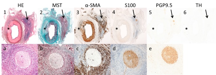
Fig 6. Nerve damage outside the lesion area.
3 weeks histology results showing a treated vessel with nerve damage outside the lesion area. A 20 x magnification (a-e) zooms in on the affected nerve that is indicated with an arrow in picture 1–6. Serial sections were stained with HE, MST, α-SMA, S100, PGP9.5 and TH. The perineurial tissue and nerves located at the opposite site of the lesion were affected by an extensive inflammatory response (1,A and 2,B), increased proliferation of myofibroblasts (3,C), a reduction in neural tissue (4,D;5,E) and loss of neurotransmitter production of the affected nerves (6).

