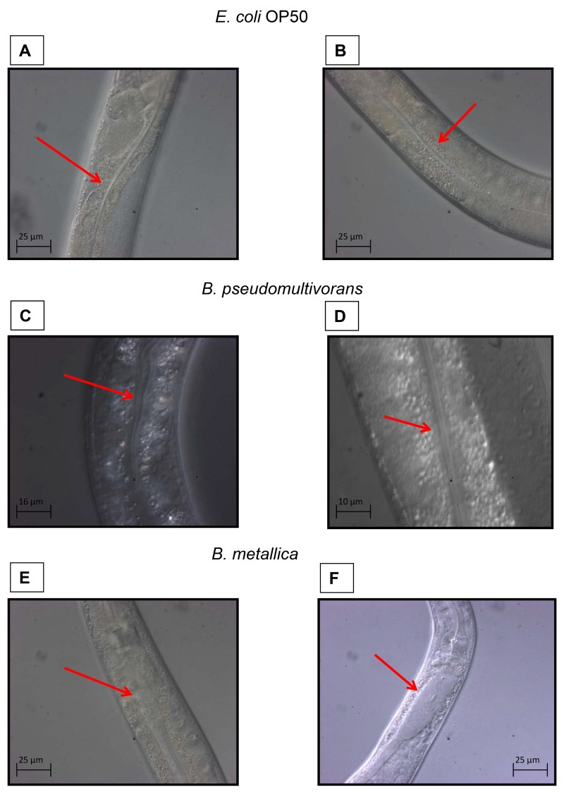
Fig 3. The ability of Bcc strains to accumulate in C. elegans intestinal lumen was evaluated with microscopy analysis.
Red arrows indicate the nematodes intestine. A) Intestinal lumen of one L4 stage WT worm after 4 h of incubation on NGM plate spotted with E. coli OP50, and B) after 24 h of incubation on the same plate. C) Intestinal lumen of one L4 WT after 4 h of incubation on NGM plate spotted with B. pseudomultivorans (VR 0 on SKA, and D) after 24 h of incubation on the same plate. E) Intestinal lumen of one L4 WT after 4 h of incubation on NGM plate spotted with B. metallica (VR 3 on SKA, and F) after 24 h of incubation on the same plate.

