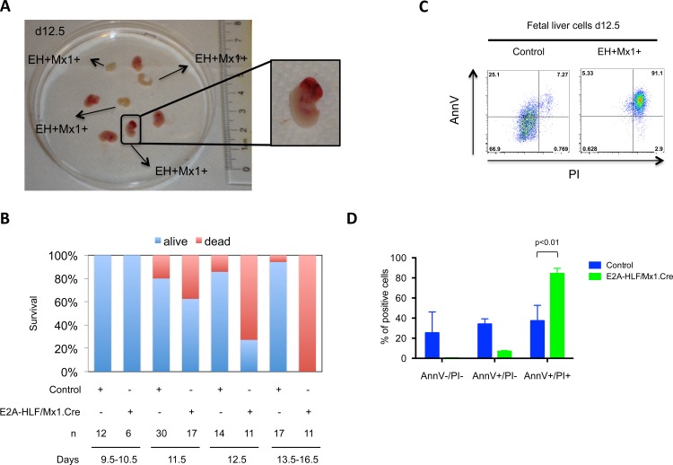
Fig 4. Expression of E2A-HLF in fetal liver hematopoietic progenitors results in embryonic lethality.
(A) Image shows embryo morphology (n = 8) at E12.5 E2A-HLF/Mx1.Cre (EH+Mx1+) embryos. An E2A-HLF/Mx1.Cre embryo in the lower part of the picture shows cranial hemorrhage and three other E2A-HLF/Mx1.Cre embryos were not viable. (B) Graph summarizing mortality of embryos depending on their genotype and developmental stage. Controls comprise wild type, E2A-HLF+/Mx1.Cre- and E2A-HLF-/Mx1.Cre+ embryos. A total of 118 embryos were analyzed. (C) Fetal liver cells of control (n = 3) and E2A-HLF/Mx1.Cre (n = 3) embryos at E11.5 were stained with annexin V (AnnV) and propidium iodide (PI) for apoptosis analysis. A representative experiment is shown. (D) Graph summarizes annexin V/propidium staining from fetal liver cells. Columns denote mean and bars denote standard error of the mean. Statistical analysis was done by Mann Whitney U test.

