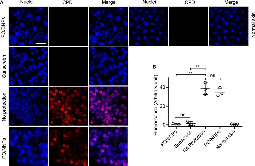
Figure 5. CPD staining of mouse dorsal epidermal sheets after receiving different topical interventions and UVB irradiation (160 J/m2).
Epidermal sheets were prepared one hour after exposure to UVB (A). The fluorescence of CPD on skin receiving different topical interventions was quantified (B). The scale bar represents 50 µm. Data are shown as mean ± SD (n=3), **p<0.01 (student t-test). Normal skin represents tissue that was not UV irradiated. BNP - bioadhesive nanoparticle, CPD - cyclobutane pyrimidine dimers, PO – padimate O.

