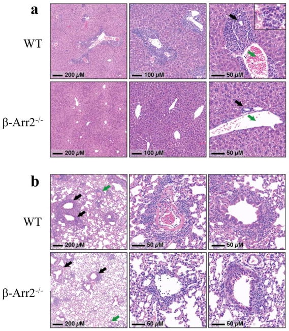
Figure 10. Loss of β-arrestin2 in T cells protects against liver and Lung damage in T cell transfer model of colitis.
Histopathology of liver and lung tissue from colitic mice 7–8 weeks post T cell transfer. (a) In the liver, inflammation was mostly confined to periportal regions (Zone 1), and consisted of a mixture of lymphocytes, eosinophils, and some neutrophils (inset, right panel). In most portal regions, inflammatory cells formed a thick rim encompassing both bile ducts (black arrow) and portal veins (green arrow). (b) Lungs from WT mice showed marked acute and chronic pneumonitis that was primarily peri-vascular (green arrows and middle panel) and peri-bronchiolar (black arrows and right-most panel) in distribution. Many of the involved parenchymal vessels showed evidence of fibrin deposition, as seen in the middle panel. Alveolar spaces were relatively spared, although significant inflammation was frequently seen extending away from vascular and airways to impinge on neighboring alveoli. Photomicrographs shown here were taken from mice with less severe, but notable inflammation that was of the same character and distribution as that seen in the WT mice.

