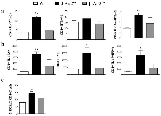
Figure 5. T cell differentiation in colitic mice.
Colonic lamina propria (cLP) cells from WT, β-arr2−/− and β-arr2+/− mice treated with DSS were stimulated ex vivo with PMA and ionomycine to determine a) proportion and b) total number of CD4+ T cells producing IL-17A, IFNγ or both cytokines. c) Cells were stained to determine proportion of RORγT+ CD4+ T cells in colons of colitic mice. N=4–7 mice for each genotype. *p<0.05, **p<0.01, p<0.001 as compared to WT using student’s t test.

