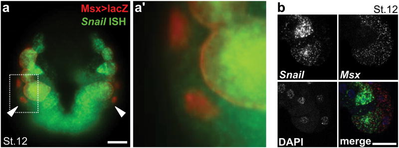
Extended Data Figure 1. In situ hybridization of neural plate border markers Snail and Msx.
a, Immunolabeling for β-Galactosidase (red) and in situ hybridization (ISH) for Snail mRNA (green) in stage 12 embryo electroporated with Msx>lacZ, revealing Snail expression in the bipolar tail neuron (BTN) progenitors (b9.36 cells, arrowheads). Dashed area enlarged in a′. b, Double ISH for Snail and Msx in stage 12 embryos counterstained with DAPI, showing co-expression in neural plate border cells, including BTN progenitors. Scale bars, 25 μm.

