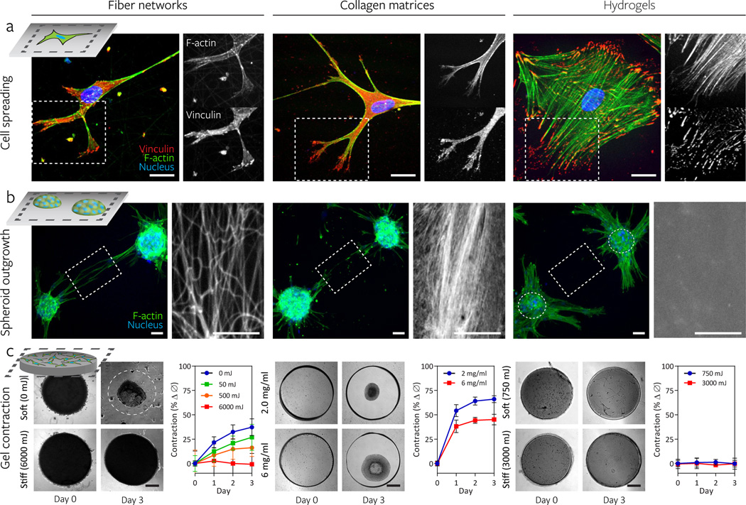
Figure 2. Synthetic fiber networks induce similar topographical and mechanical interactions with cells as collagen matrices at multiple length scales.
Comparison of cell spreading (a), spheroid outgrowth (b), and gel contraction (c) across hMSC-seeded DexMA fiber networks (left column), type I collagen matrices (middle column), and flat DexMA hydrogels (right column). a, Cytosolic and focal adhesion-localized vinculin (red), counter-stained for F-actin (green) and cell nuclei (blue) with phalloidin and Hoechst 33342, respectively. Dashed box designates region shown with F-actin or vinculin channel isolated (scale bars, 50 µm). b, Outgrowth of hMSC spheroids (100 cells/spheroid) stained for F-actin (green) and cell nuclei (blue). Rhodamine methacrylate-coupled DexMA fibers and hydrogel surface and Picrosirius Red-stained collagen are shown in the region spanning two spheroids, as indicated by the dashed box (scale bars, 50 µm). c, DexMA fibers and hydrogels and collagen were processed into thick circular slabs and seeded at high density with hMSCs. Initial and final states of soft (top) and stiff (bottom) constructs. Cell-mediated contraction was determined by normalizing construct diameter to the initial diameter; mean ± s.d., n ≥ 6 (scale bars, 500 µm).

